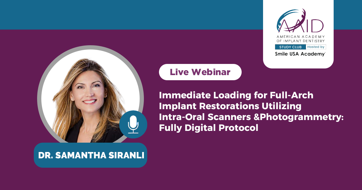
Immediate Loading for Full-Arch Implant Restorations Utilizing Intra-Oral Scanners and Photogrammetry : Fully Digital Protocol
A fully digital approach to immediate full-arch implant restoration using intraoral scanning and photogrammetry for precise, same-day results.

with Dr. Samantha Siranli
Panelists
1 expert lined up.
About this webinar
This fully digital protocol demonstrates how the integration of facial scanning, intraoral scanning, photogrammetry, and in-house milling can enhance efficiency, improve accuracy, and provide predictable immediate full-arch implant rehabilitation.
Our protocol begins with comprehensive digital records obtained prior to surgery, including extra-oral 3-D facial scans, 2-D photographs, and intraoral scans. These records allow for precise digital planning of tooth position, smile design, and the horizontal and vertical dimensions of occlusion. Multiple clinical cases will be presented in detail to demonstrate the digital smile design process, the merging of the initial intraoral scans with the newly designed smile, and the transfer of this design by overlapping it with the post-surgical scan bodies and photogrammetry data following implant placement.
By integrating these technologies, the workflow allows the predictable fabrication and delivery of a milled PMMA full-arch provisional prosthesis on the day of surgery, providing patients with immediate esthetics, function, and a fully guided occlusal scheme.
Lecture Objectives
• Step-by-step clinical demonstration of the complete digital workflow
• Merging facial scans with intraoral scans for smile design and determination of the vertical dimension of occlusion prior to surgery
• Intraoral scanning of abutments following implant placement
• Photogrammetry scanning of abutments for precise implant position capture
• Merging abutment scans with the initial intraoral scans using anatomical references or half scan technique
• Computer-aided design and computer-aided manufacturing (CAD/CAM) of the provisional prosthesis
• Final finishing and adjustment of the milled PMMA prosthesis prior to insertion on the day of surgery
-
Date
Sun, 12 Apr 2026
-
Time
7:30 PM IST
-
CE credits
1 hours
-
Content Format
Live webinar
-
Access
Live + Recorded · Recording available afterwards
-
Certificate
Issued after attendance
-
Platform
Zoom Premium Webinar
-
Hosted by
Smile USA & Dentist Channel
Related webinars

Principles of Modern Exodontia – Atraumatic Extractions and Alveolar Bone Preservation!
Master modern exodontia with advanced techniques in atraumatic extractions and alveolar bone preservation in this expert...

The Role of the modern dentist in treating Edentulous sites
Discover the role of modern dentists in treating edentulous sites using advanced techniques in this comprehensive webina...
 Free
Free
Practical solutions to manage implant complications

Alveolar Ridge Deficiency: Surgical Algorithm, Treatment Considerations
Explore surgical algorithms and treatment considerations for managing alveolar ridge deficiency in implant dentistry.
